
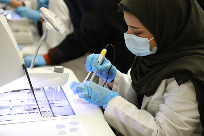
Training & Education
Nourah’s Tissue Biobank training program aims to help health researchers and graduate students to explore biobank topics and applications. Nourah’s Tissue Biobank consists of three main units, Histopathology, Cytopathology and Advanced imaging unit. In addition, we collaborate with genetic section laboratories located at the Health Science Center to perform DNA and RNA isolation, PCR and DNA sequencing.
The Training Program
- A program will be constructed based on the qualifications and needs of the trainee.
- The program objectives, a schedule of training sessions and periodic evaluations of the trainee will be handled by the training instructor in each specialization.
Qualifications required to be able to enroll in Nourah’s Tissue Biobank training programs:
- Health-related specialists
- Physics and radiology
- Other specialists interested in a career in health sciences research
- Applicants in their final year of an undergraduate program, internship, or postgraduate trainee
To apply for Nourah’s Tissue Biobank training program
Nourah’s Tissue Biobank Learning Topics
Nourah’s Tissue Biobank training program is designed for any health researchers and graduated students who are interested to learn more about biobank or any of the scientific applications we use in histopathology, cytopathology and advanced imaging units. Training program will include theory and practical skills.
Theoretical education is designed to include general information regarding Tissue Biobank such as:
The ethical issues associated with the use of biospecimens in research
Regulatory requirements that will be complied with ISO accreditation
Tissue and information release (material release)
Material handling (tissue and information processing and storage)
Facility security and procedures
Maintaining records and updating inventories and databases
The importance of biobanks in health research
Technical Training and processes relevant to operations at the site
Histopathology Unit
You will learn about different types of human tissues received in Nourah’s Tissue Biobank. You will go through tissue processing, embedding, cutting and staining techniques
Cytopathology Unit
You will learn about different types of cells received in our unit and how to stain and process them at the cellular level
Advanced Imaging Unit
This unit is divided into three different imaging techniques including:
-
Electron microscope
- Scanning electron microscope (SEM): SEM produces image of a sample by scanning the surface with a focused beam of electrons. The electrons interact with atoms in the sample, producing various signals that contain information about the surface topography and composition of the sample.
- Transmission electron microscope (TEM): Transmission Electron Microscope works when a beam of electrons is transmitted through a specimen to form an image. An image is formed from the interaction of the electrons with the sample as the beam is transmitted through the specimen.
-
Confocal microscopy
Confocal microscopy is a technique that uses lasers and fluorescence to create a three-dimensional image of a sample. A focused laser beam is used to excite the molecules at one point of the sample. The molecules, called fluorophores, release photons as they return to their unexcited state, causing the fluorescence
-
Micro CT Skyscan.
MICRO CT Skyscan 3D X-ray systems offers solutions for non-destructive, high resolution 3D imaging of small laboratory animals and a wide variety biological sample